Nursing Diagnoses:
1. Compromised airway r/t altered LOC evidenced by fluctuating GCS levels within the past 24 hours.
Evidence:
- Fluctuating GCS of 9-11
- Tracheotomy
- Occasional Restraints to prevent pulling of IV
- Confusion
- Bedrest
- Thick creamy secretions
- Diet: Full thickened fluids
- Patient risk for aspiration.
Interventions
- Apply suction prn to maintain clear airway when secretions are present.
- Administer analgesics to maintain patient comfort (shallow breathing can be induced by pain d/t discomfort)
- Encourage deep breathing and coughing
- Hyperoxygenate before and after tracheal suction
- Maintain patient hydration and encourage fluids to liquefy thick secretions
- Assess patient oxygen saturation and respiratory rate, assess present oxygen therapy devices.
- Auscultate breath sounds and assess air movement to monitor and record status.
- Observe for signs of respiratory distress (increased rate, restlessness/anxiety, use of accessory muscles for breathing)
- Observe for improvement in symptoms
- Contact respiratory therapy to assess the patient’s respiratory situation
- Monitor the blood gas values
- Educate the patient and family on why he has a tracheotomy, what your monitoring for and answer any questions they might have
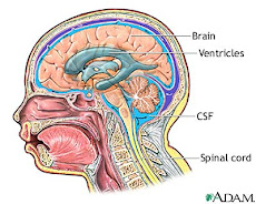
2. Altered cerebral tissue perfusion r/t IVH and EVD placement.
Evidence:
- EVD-draining cloudy red CSF
- Hydrocephalus
- Increased Intracranial pressure
Fluctuating GCS - Aphasia
- Decreased ADL’s
- Confusion
- Bedrest
- Right sided weakness
- Atrial Fibrillation
- Discontinued anticoagulant therapy
- Bowel and Bladder Incontinence
Interventions
- Neuro vitals q2h
- Assess for purposeful and non-purposeful motor responses, compare right and left sides.
- Decrease extraneous stimuli/provide comfort measures (ex. Quiet environment and rest) to reduce CNS stimulation.
- Monitor/document changes in LOC and indications of increased ICP (pupils, headache, numbness/tingling).
- Monitor vital signs
- Provide safety measures to prevent injury (side rails, assess restraints frequently)
- Assess the patient’s pain level on a scale of 0-10, administer analgesics prn
- Educate the patient and family on IVD’s & EVD’s, inform them what you are monitoring for and answer any questions they might have
3. Tissue Injury r/t traumatic catheter insertion evidenced by gross hematuria.
Evidence:
- Gross hematuria
- 3 way Foley Catheter inserted
- Discontinued anticoagulant therapy
- Previously high INR
- Cerebral hemorrhage
Interventions
- Investigate urology consult.
- Assess I&O and characteristics of output (amount of hematuria, presence of clots), identify if hematuria has decreased or increased. Document findings.
- Assess signs of bleeding in other areas (nosebleeds, signs of increased ICP, bruising)
- Administer analgesic medication to improve client comfort and relieve pain.
- Assess CBC values for evidence of increased bleeding (decreased RBC, decreased Hemoglobin etc)
- Monitor INR and PTT closely
- Assess skin color and cap refill.
Assess Vitals signs q4h (note BP and HR) - Provide safety measures to prevent injury (side rails, check restraints)
- Educate the patient and family on why blood is in his urine, what you’re monitoring for and answer any questions they might have
Schedule:
Patient Name: Wilfred Longbottom Age: 82 years old Room and Bed #: Obs 2 Bed 3
0800
- Meds (1,2,3), VS (1,2,3), Neuro vitals (1,2)
- Initial Assessment (complete head to toe) (1,2,3), Assess pain-give pain meds prn (1,2,3)
- Check equipment including oxygen therapy devices and side rails (1,2,3)
- Check IV NS @ 120ml/h
- Review I&O sheet from evening shift (3)
- Assess the characteristics of the output (3)
0900
- Encourage DB & C (1)
- Assist with breakfast (1)
- Basin Bath
- Check IV NS @120 ml/h
1000
- Meds (1,2,3)
- Interpret blood gases and take necessary action (1)
- Read lab value & take necessary action (1,2,3)
- Chem Strip
- Check IV NS@120ml/hr
- My break
1100
- Charting (1,2,3)
- Patient and family teaching (2)
- Check IV NS@120ml/hr
1200
- Meds
- VS (1,2,3), Neuro Vitals (1,2)
- Assess pain level (1,2,3)
- Assist with lunch(1)
- Check IV NS@120ml/hr
1300
- Check IV NS@120ml/hr
- My Lunch
1400
- Empty Catheter, record I&O & assess output (3)
- Chem Strip
- Check IV NS@120ml/hr
- Allow the patient a quiet environment to rest (2)
1500
- Check IV NS@120ml/hr
- Patient & family teaching (1,3)
1600
- Vital Signs (1,2,3)
- Neuro Signs (1,2)
- Check IV NS@120ml/hr
1700
- Meds (1,2,3)
- Check IV NS@120ml/hr
- Assist with supper (1)
- My break
1800
- Review charting (1,2,3)
- Chem Strip
- Check IV NS@120ml/hr
- Empty catheter, record I&O & assess output(3)
1900
- Check IV NS@120ml/hr
- Report off
- Report
*Apply suction prn (1)
Head-To-Toe Assessment
Nursing Diagnosis
1. Compromised airway r/t altered LOC evidenced by fluctuating GCS levels within the past 24 hours.
2. Altered cerebral tissue perfusion r/t IVH and EVD placement.
3. Tissue Injury r/t traumatic catheter insertion evidenced by gross hematuria.
- Glasgow Coma Scale (1,2)
- Level of Conciousness (1,2)
- Level of Orientation (person, place & time) (1,2,)Ask about level of pain on a scale of 0-10, 0 being no pain at all and 10 being the worst pain possible. Ask where the pain is, describe what type of pain it is, how long the pain has continued for and anything that brought on the pain (PQRST method). Administer pain meds as neccessary. (1,2,3)
- Pupillary Reflex- assessing whether they are equal, brisk or sluggish and what size (1,2)
- Activity and Movement-check for movement of all limbs and strength of handgrips, arms, legs, plantar flexion and dorsiflexion. Rate the strength of each limb according to weak, moderate and strong and if its equal bilaterally. Take note of the patient’s activity level which in this case is bedrest. (2)
- Sensation-ask if the patient is experiencing any feelings of numbness or tingling. Identify where its locate and if the feeling radiates elsewhere. (2)
- EVD- note the amount, color and consistency of the fluid draining from the EVD. Look for any present blood clots. (2)
- Color (pallor, erythema, cyanosis, jaundice or congruent with skin type). Assess the body for skin color throughout, pay special attention to the nailbeds and lips for cyanosis which would indicate respiratory distress. (1,2)
- Temperature-take an oral temperature. Feel for skin temperature with the dorsal side of your hands over all areas of the body,comparing symmetric spots. (1,2,3)
- Perpheral Pulses- carotid, radial, brachial, popliteal, dorsalis pedis and posterior tibial pulses. Rate each pulse from 0-4 and compare bilaterally. Also check cap refill and skin turgor. (2)
- Edema- Look for any edema throughout the extremities. If any pitting edema is noted grade it from +1-4.
- Heart sounds- inspect, palpate, percuss and auscultate the heart
- Vital Signs- TPR, BP & O2 sats (1,2,3)
- SOB/SOBOE- observe/ask the patient if they feel short of breath. (1)
- Respiratory rate- assess the rhythm, rate, the position they take to breath and any use of accessory muscles.(1)
- Trach secretions-note the amount, consistency and color of the secretions. Apply suction as necessary. (1)
- Cough- is it productive or not. Observe whether the client is able to clear their own secretions. (1)
- Breath sounds-inspect, palpitation, percussion, auscultate lund fields. (1)
- O2 sats- assess if the client needs additional oxygen. (1)
- Diet- how well the patient tolerates their fully thickened fluid diet by observing them eatting. Ask them if they have an appetite. (1)
- Nausea/Vomiting- is the patient experiencing any nausea. Adminster gravol as neccessary. (1,2,3)
- Abdomen- inspect, auscultate, percuss and palpate the abdomen. Note if the abdomen appears scaphoid, distented, rounded or flat. Listen for bowel sounds in all four quadrants. (3)
- Catheter- assess the I & O ratio, color, amount and consistency of the urine. If gross hematuria continues, monitor it closesly throughout the day. (3)
- Bowel/Bladder patterns- take note of the patients last bowel movement. When changing the benefits look for incontinence of stool.
- Skin condition- observe the skin for temperature, moisture, texture, thickness, bruising, and lesions. (2,3)
- Dressings-inspect the dressing for any drainage. Lightly palpate over the dressing for any lumps. (2)
- Inspect the groining for tissue breakdown related to catheter insertion (3)
- Coping-ask the patient how he’s coping with being in the hospital, reduced verbal communication and the change to his present health status.
- Visitors-ask if he will have vistors in to see him.
- Mood/Affect- ask about the patient’s mood and monitor if it’s congruent with his affect.
Neuro Vital Signs
Nervous System Assessment
- Assessment if mental status, cranial nerves, cerebellar function, sensory & motor system and reflexes.
Neuro Vital Signs/
- Assess intracranial activity by looking at level of consciousness (LOC), vital signs, pupilary reaction, and motor responses.
- Score ranges from 3-15.
- Other considerations that could alter assessment: intubated, eyes swollen closed, paralyzed, doesn’t speak English, deaf, aphasic, blind.
- detects life-threatening complications such as cerebral edema
- How would you assess infant/toddler? (crying, playing, laughter)
a. Level of Consciousness
- first change with altered cerebral tissue perfusion
1. Response to touch-
- call patient by name before touch and ask to open eyes
- if no response, repeat stimulus in both ears, loudly
- If no response, touch patient firmly and repeat command. If no response, notify RN
2. Orientation
- Person (Please tell me you name/ what’s your name?)
- Place (What building are you in? City? Province?)
- Time (What is today’s date? Day of the week? Month? Year? Season?)
B. Pupils
- eye opening
- talk and observe response
- avoid direct light
- estimate pupil size(
- Use flashlight from periphery of eye to determine response (
- Repeat on other eye (check for bilateral/unilateral concerns)
C. Motor Response
- Increased ICP at cortical levels leads to abnormal flexion posturing. As it increase at pons, it leads to abnormal extension posturing. At medulla there is flaccidity. No response is worst.
- Upper limbs
- ask client to move limbs
- Use 2 fingers and ask client to grab them firmly.
- ask client to release grip ( grip may be reflex, release is cognitive)
2. Lower Limbs
§ ask client to move limbs
§ ask client to push against resistance ( test bilateral at same time)
D. Sensory response
1. Have client close eyes
2. Touch client on all four limbs and ask to identify touch
E. Vital Signs
1. Take pulse, resps, BP, and report changes
2. These would be late changes
3. Record all data
Cushing’s response-
- Bradycardia, systolic hypertension, wide pulse pressure and alterations in respiratory response (bradycardia, low resp rate and wide pulse pressure is Cushing’s triad)
- Due to pressure on medulla
Signs of Increased ICP
- headache
- nausea/vomiting
- seizures
- irritability
- drowsiness
- decreased activity
- increased fatigue
- memory loss
- late signs: decreased LOC, response, alterations in pupils
Coma occurs because of extensive, diffuse, bilateral cerebral hemispheric destruction, a lesion in diencephalons, destruction of brainstem down to level of pons.